In short
- Marrow mesenchymal stem cells can become bone-forming cells or fat cells. With age, the balance tips toward fat.
- In human hip-bone biopsies, older age went with more marrow fat and less trabecular bone, and the two were inversely related.
- In a 2025 study, old male mice given PEMF for four weeks formed more bone and less marrow fat. Removing sensory nerves abolished the change.
- It was an animal study with about five mice per group, and young mice showed no change. It cannot be translated into conclusions about human bone health.
Bone or fat?
Bone is not finished once it is built. Osteoblasts lay down new bone and osteoclasts remove old bone throughout life. One source of new osteoblasts is the mesenchymal stem cells in the marrow, which can become either osteoblasts or fat cells. You can explore the skeleton in our body atlas.
A 2004 study in Aging Cell by Moerman and colleagues reported that with age, fewer marrow stem cells commit to the osteoblast lineage and more to the fat lineage. Bone-building genes such as Runx2 were expressed less, while PPAR-γ2, which drives fat-cell development, was expressed more.
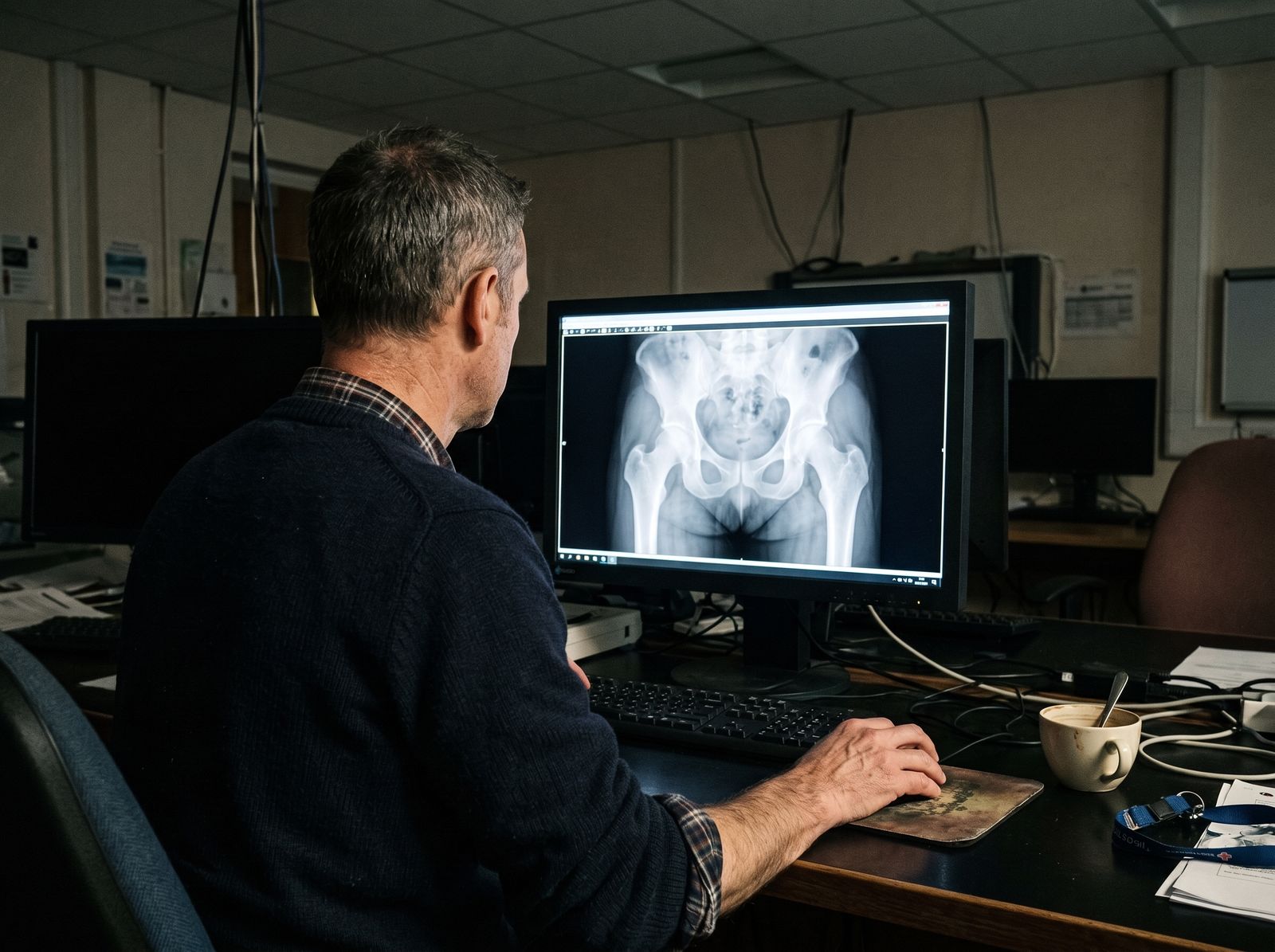
People show a similar pattern. In 2001 Danish researchers analysed hip-bone biopsies from 53 healthy adults aged 30 to 100 and 26 people with osteoporosis. With age, the share of marrow taken up by fat rose and the share of trabecular bone fell, and the two were inversely correlated. People with osteoporosis had more marrow fat than age-matched controls. This is a correlation, and the authors noted the mechanism was still unknown.
Sensory nerves inside bone
Bone contains nerves as well as blood vessels. A 2013 mouse study in Nature by Fukuda and colleagues showed that a protein made by nerve cells, semaphorin 3A (Sema3A), is needed to maintain bone mass. Mice lacking Sema3A only in neurons had fewer sensory nerves in their bone and lower bone mass, and removing sensory nerves in normal mice also reduced bone mass.
In mice, then, sensory nerves are not just pain wiring; they also send signals that take part in building bone.
What the 2025 PEMF study did
PEMF (pulsed electromagnetic fields) uses a magnetic field switched on and off in short bursts to induce weak currents in tissue; the principle is covered in PEMF and whole-body vibration. In a 2025 study in Nature Communications, Wang and colleagues exposed young (4-month) and old (20-month) male mice to PEMF at 8Hz and 3.8mT for one hour a day over four weeks.
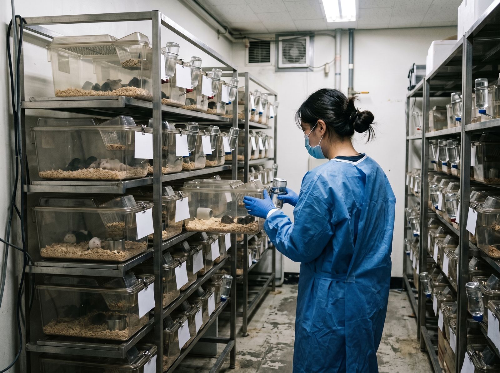
- In old mice, trabecular bone volume fraction and trabecular thickness in the thigh bone increased, along with a marker of bone formation. Young mice showed no such change.
- Marrow stem cells expressed fewer fat-differentiation genes, and bone-resorbing osteoclasts were unaffected.
- In mice whose sensory nerves were genetically removed, PEMF no longer increased bone formation.
- Sensory neurons exposed to PEMF secreted more Sema3A, and blocking its receptor Nrp1 on the stem cells cancelled the effect.
The core finding is that in this mouse model a physical stimulus from outside the body was linked, via sensory nerves, to the direction stem cells take.
What this study does not say
The limits are clear. Only male mice were studied, with key measurements on about five animals per group. One frequency and intensity was tested for four weeks, and young mice did not respond. The authors themselves note that the amount of new bone seen in mice may differ in human patients. The study does not address females, other settings, or bone density and fracture risk in people.
Human PEMF research has mostly looked at fractures that fail to heal. A 2011 Cochrane review of four randomised trials (125 people) concluded that electromagnetic stimulation may offer some benefit, but the evidence was inconclusive and insufficient to guide practice.
If you are worried about your bones
- If osteoporosis or fractures concern you, start with a bone density test and a doctor’s assessment; diagnosis and treatment belong with your doctor.
- If you have a pacemaker, implantable defibrillator, cochlear implant or other implanted electronic device, talk to a doctor before using any PEMF device.
- If you are pregnant or being treated for a condition, check with your doctor first.
- PEMF does not replace diagnosis or treatment.
BAHIOLOGY and Wave Motion
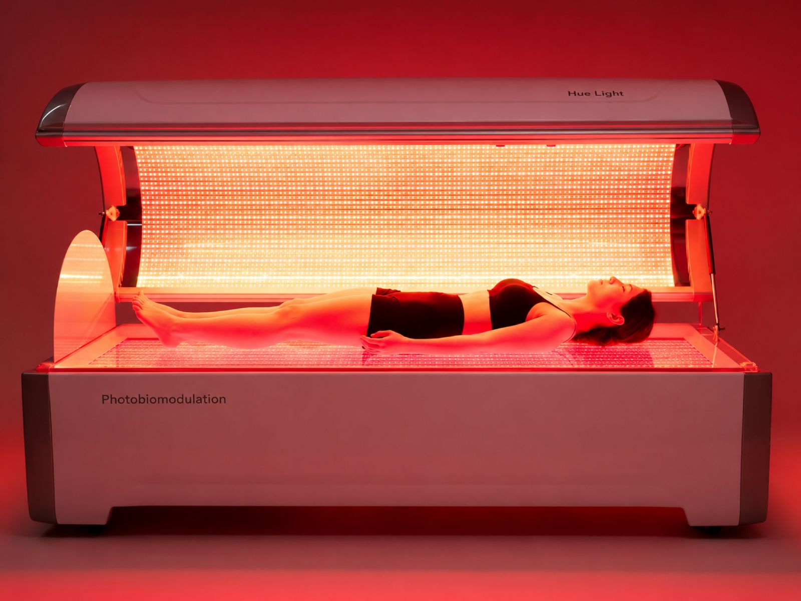
BAHIOLOGY’s Wave Motion delivers PEMF with adjustable frequency and intensity together with low-frequency waves and vibration; BAHIOLOGY researches and develops it, built together with its investor and manufacturing partner Hue Light Co., Ltd. You can see it in four technologies. This article explains research findings; it does not promise any particular effect.
Questions
Why does bone marrow fat increase with age?
Marrow mesenchymal stem cells can become bone-forming cells or fat cells, and research suggests a larger share heads toward fat with age. The exact mechanism is still being studied.
Does PEMF help osteoporosis?
The 2025 study observed changes in bone formation in old male mice. It cannot tell us whether PEMF helps osteoporosis in people, and osteoporosis needs a doctor’s diagnosis and treatment.
How is PEMF thought to affect bone?
In this mouse study, PEMF stimulated sensory nerves in bone to release more Sema3A, a signal that pushed stem cells toward bone. Whether the same pathway works in people is unknown.
Sources
- Wang T, et al. Pulsed electromagnetic fields mediate sensory nerve regulation for bone formation in aging models. Nat Commun. 2025
- Moerman EJ, Teng K, Lipschitz DA, Lecka-Czernik B. Aging activates adipogenic and suppresses osteogenic programs in mesenchymal marrow stroma/stem cells: the role of PPAR-gamma2 transcription factor and TGF-beta/BMP signaling pathways. Aging Cell. 2004
- Justesen J, et al. Adipocyte tissue volume in bone marrow is increased with aging and in patients with osteoporosis. Biogerontology. 2001
- Fukuda T, et al. Sema3A regulates bone-mass accrual through sensory innervations. Nature. 2013
- Griffin XL, Costa ML, Parsons N, Smith N. Electromagnetic field stimulation for treating delayed union or non-union of long bone fractures in adults. Cochrane Database Syst Rev. 2011
Want to know more?
If you’d like to try BAHI Longevity or talk to us, leave your details and we’ll be in touch.